What is optical coherence tomography of the eye?
Optical coherence tomography is a non-invasive( noncontact) method for studying the structure of the eye tissue. It allows you to get pictures of a higher resolution than the results of ultrasound procedures. In fact, optical coherence tomography of the eye is a type of biopsy, only for carrying out the first there is no need to take a tissue sample.
A short digression into the history of
The concept, based on which modern optical coherence tomography is performed, was developed by researchers at the Massachusetts Institute of Technology in the distant 1980s. In turn, the idea of introducing a new principle into ophthalmology was proposed in 1995 by the American scientist Carmen Puliathito. A few years later, Carl Zeiss Meditec developed a suitable instrument called Stratus OCT.
Currently, with the help of the latest model, it is possible not only to study retinal tissues, but also optical coherence tomography of the coronary arteries, optic nerve at the microscopic level.
Principles of the
study Optical coherence tomography is the formation of graphic images based on the measurement of the delay period when the light beam reflects from the tissues under examination. The main element of the devices in this category is a superluminescent diode, the use of which makes it possible to form light beams of low coherence. In other words, when the apparatus is activated, a beam of charged electrons is divided into several parts. One stream is directed to the area of the tissue structure under study, the other to the special mirror.
The rays reflected from the objects are summarized. Subsequently, the data are recorded by a special photodetector. The information formed on the graph allows the diagnostician to draw conclusions about the reflectivity in individual points of the object under study. When evaluating the next section of the fabric, the support is moved to another position.
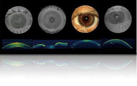
Optical coherent tomography of the retina makes it possible to generate graphs on the computer monitor, which are in many ways similar to the results of ultrasound examination.
Indications for the procedure
Today, to make optical coherence tomography is recommended in the diagnosis of such pathologies as:
- Glaucoma.
- Macular tissue ruptures.
- Thrombosis of Retinal Veins.
- Diabetic retinopathy.
- Degenerative processes in the structure of the eye tissue.
- Cystoid edema.
- Anomalies in the functioning of the optic nerve.
In addition, optical coherence tomography of the optic nerve is assigned to evaluate the effectiveness of the therapeutic procedures used. In particular, the method of investigation is indispensable in determining the quality of the installation of the drainage device, which is integrated into the eye tissue for glaucoma.
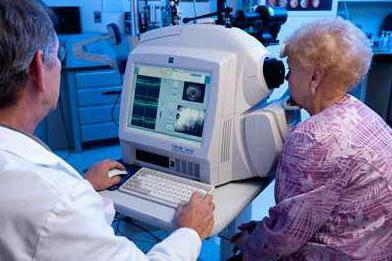
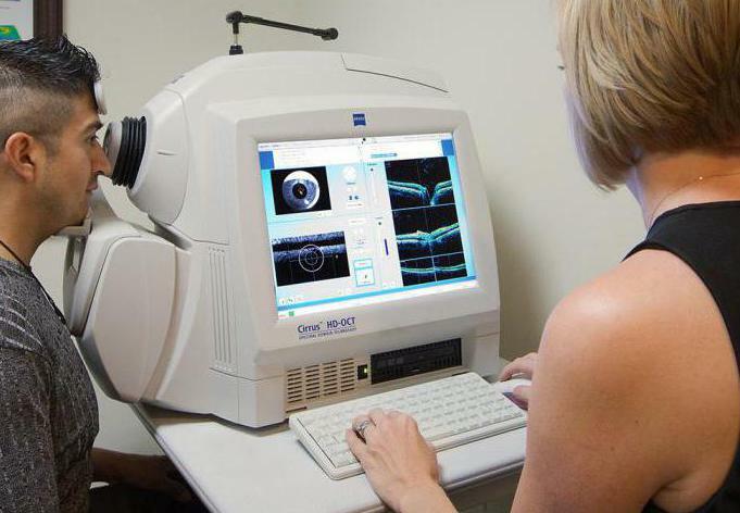
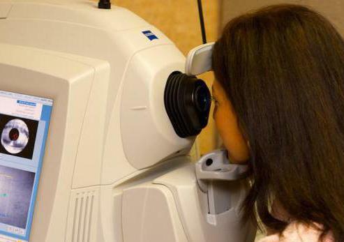
Diagnostics features
Optical coherence tomography imposes a focus on the subject's vision on special marks. In this case, the operator of the device performs a series of consecutive scans of tissues.
Such pathological processes as corneal edema, profuse hemorrhages, all kinds of opacities are able to impede the investigation and prevent effective diagnosis.
The results of coherent tomography are formed in the form of protocols that inform the researcher about the state of certain tissue sites, both visually and quantitatively. Since the obtained data are fixed in the memory of the device, they can subsequently be used to compare the state of the tissues before the treatment and after the application of the therapy methods.
Three-dimensional visualization of
Modern optical coherence tomography makes it possible to obtain not only two-dimensional graphics, but also produce three-dimensional visualization of the objects under study. Scanning of tissue sites at high speed allows the formation of more than 50,000 images of the diagnosed material within a few seconds. Based on the received information, special software reproduces on the monitor the volume structure of the object.
The generated 3D image is the basis for the study of the internal topography of the eye tissue. Thus, it opens the possibility to define clear boundaries of pathological neoplasms, as well as to fix the dynamics of their changes in time.
Advantages of coherent tomography
The most effective coherent tomography devices are demonstrated in the diagnosis of glaucoma. In the case of using devices of this category, specialists are able to accurately determine the factors of pathology development in the early stages, to reveal the degree of progression of the disease.
The method of investigation is indispensable in diagnosing a common disease such as tissue masculodystrophy, in which, as a result of the age-related characteristics of the body, the patient begins to see a black spot in the central part of the eye.
Coherent tomography is effective in combination with other diagnostic procedures, for example, with angiography of the retina in a fluorescent way. When the procedures are combined, the researcher receives especially valuable data, which contributes to the correct diagnosis, the complexity of the pathology and the choice of effective treatment.
Where can I perform optical coherence tomography?
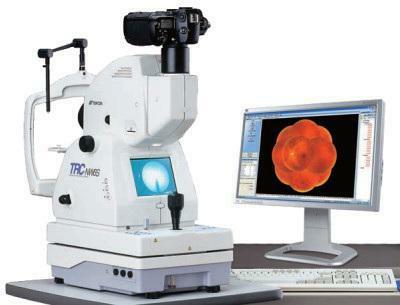
The procedure is possible only if there is a specialized OCT device. Diagnostics of such a plan can be resorted to in modern research centers. Most often, such equipment is equipped with vision correction cabinets, private ophthalmology clinics.
Question price
Conducting coherent tomography does not require the direction of the attending physician, but even with its presence, diagnostics will always be paid. The cost of the study determines the nature of the pathology, the detection of which is aimed at diagnosis. For example, the definition of macular tissue ruptures is estimated at 600-700 rubles. While carrying out a tomography scan of the anterior part of the eye can cost the patient of the diagnostic center 800 rubles or more.
As for complex studies aimed at assessing the functioning of the optic nerve, the state of retinal fibers, the formation of a three-dimensional model of the visual organ, the price for similar services today starts from 1800 rubles.