How is MRI decoded in medicine?
This method of examination is widely used in modern medicine. MRI helps detect pathological changes in many organs. For those who do not know, as the MRI stands for - is Magnetic Resonance Imaging. Let us consider in more detail what the procedure is and why it is used.
MRI - the word
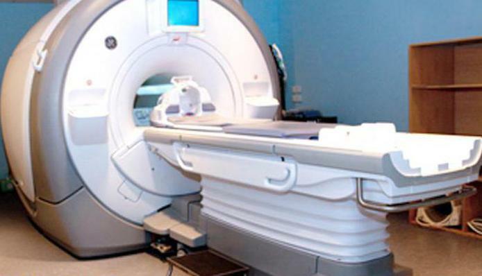
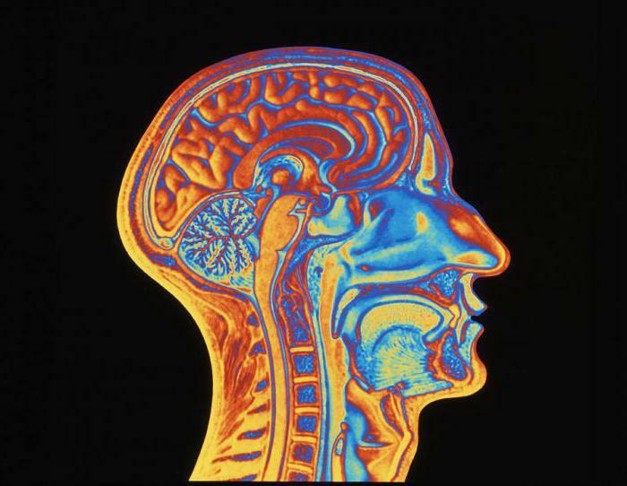
MRI( Magnetic Resonance Tomography) is a modern high-tech non-radial method used to diagnose various pathologies of tissues and internal organs.
Without resorting to internal intervention, with MRI, it is possible to obtain a highly informative image of any body structures, including volumetric projections.
The advantage of the survey is its complete safety, ionizing radiation is completely excluded. Diagnosis is carried out even by pregnant women and young children.
Medicine has a wide range of diagnostic capabilities through the use of MRI.For the result to be qualitative, during the examination the patient must strictly follow the directions of the doctor
. The MRI stands for , knows the specialist, he takes into account the data of other studies and anamnesis. 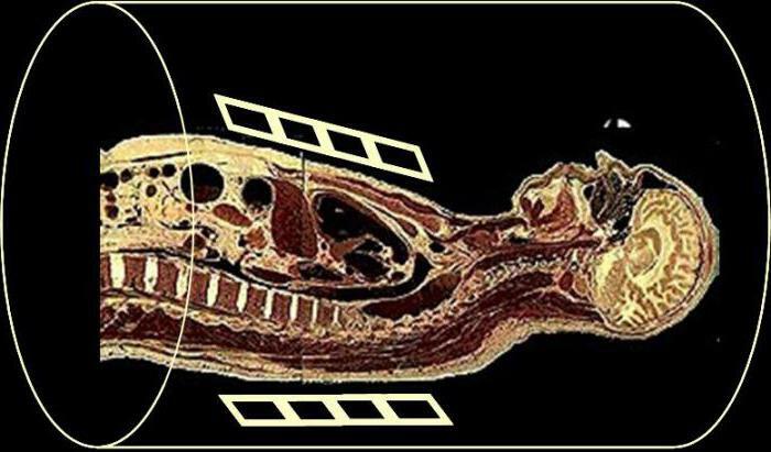
Basic indications for MRI
The following conditions are recommended for examination on the MRI device:
-
and blood circulation abnormalities;
-
defects in development;
-
various tumor processes;
-
follow-up of a patient after surgery or during therapy;
-
degenerative, demyelinating changes;
-
arterio-venous malformation , aneurysm, thrombosis, stenosis of vessels, various vascular pathologies;
-
inflammatory processes;
-
chronic, acute sinusitis;
-
suffered trauma;
-
arthrosis, bursitis, arthritis;
-
compression of the spinal cord, roots of the spinal nerves;
-
heart disease, ischemic disease, myocardial infarction;
-
damage to tendons, ligaments, joints, meniscuses, nerve endings, muscles;
-
pathology of the pelvis, abdominal organs.
If the doctor reveals from the patient as , the of the listed conditions, he sends the patient to the MRI for a reliable examination. After
as the MRI is decoded, the is established with an accurate diagnosis.
Advantages of MRI
In medicine, the advantage of MRI is given in the diagnosis of soft tissue pathologies. A wide application of the survey was received in oncology, in the diagnosis of diseases of the brain, spine, as well as in angiology and some other fields of medicine. The key advantages of this method are: the
-
does not have a radial load when compared with a CT scan;
-
method highly informative with diagnosis of tumor development in the early stages;
-
high-quality image possible without the use of contrast ;
-
examination clarifies not only the structure, but also the functional parameters ( blood flow velocity, cerebrospinal fluid, activation of the cerebral cortex). How the MRI is deciphered for these parameters, the trained specialist knows.
MRI is practically not used to diagnose pathologies of the lungs, intestines, stomach and bones.
How the
is being tested Special preparation for MRI is not required in most cases. An exception is the MRI of the abdominal cavity.
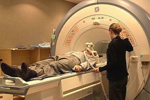
Before passing the procedure, the doctor asks the patient to remove all items from the metal( buttons, jewelry, etc.), as they will negatively affect the quality of the examination.
The patient passes into the research room, where he is placed in a special tube. In some devices during the study you can stand, but the image quality at the same time is lower.
During the procedure, it is possible to talk to the doctor through the intercom device, he closely follows the patient with video equipment.
For maximum quality image, the main requirement is complete immobility. The process takes about half an hour.
Contrasting agents may be used to clarify certain areas.
Some patients are disturbed by noise, this is the norm in the work of the technique. To eliminate discomfort, you can use headphones.
Some experience claustrophobia, such patients are recommended to conduct an examination in an open-type apparatus.
If a test is performed in newborns, young children, the doctor uses short-term anesthesia, because it is difficult for babies to remain immobile.
Those who are worried before the procedure, have any negative experience, can take easy sedatives for calming.
Contraindications MRI
Although MRI examination is a fairly safe procedure, still has contraindications:
-
the patient has a pacemaker;
-
metal plates, any splinters, apparatus Ilizarov ;
-
some implants in the middle ear;
-
is not recommended for examination in patients with unstable psyche;
-
pregnant in the first trimester - there is still no information about how the magnetic fields affect the development of the fetus;
-
coma or severe illness( decompensation stage);
-
the presence of tattoos if any dyes based on metals are contained.
-
if a contrast medium is used, allergic reactions to constituent components are also contraindications.
How MRI is deciphered in medicine
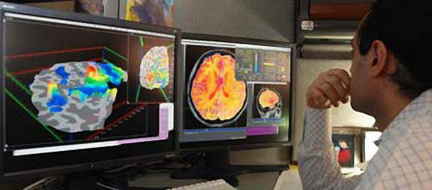
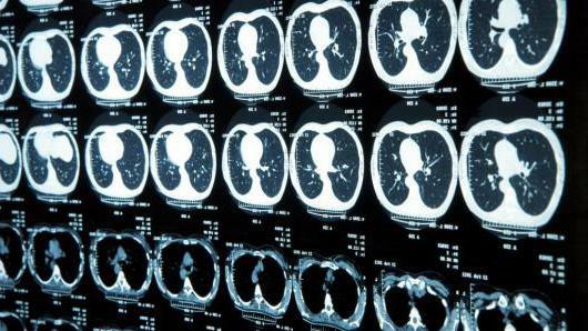
The results of the MRI scan are in the form of layered images. They can be immediately seen on the monitor, they are recorded on a disc that the patient receives along with the pictures.
The main factor of diagnostics is the interpretation of the results. This is not an easy process, which requires special knowledge of . How to decode the MRI of the brain or of another organ, only a professional knows.
A correct interpretation of the result together with other indicators from the medical history enables the doctor to establish an accurate diagnosis and to prescribe the correct effective treatment.
In some cases, for the interpretation of MRI results, some patients receive a remote consultation via the Internet.
If the case is complex, controversial, the doctor makes several images in different angles, so the picture becomes more complete and reliable . The diagnosis is set more precisely.
As for the length of the decryption, it takes much longer than the survey itself. For example, MRI of the brain is decoded about an hour and a half.
Is it possible to decode the
MRI itself is the most accurate method in diagnosis. The patient receives black-and-white photographs on his hands, as well as records on digital media. In addition, with the pictures issued a conclusion of specialists, which is written on the basis of the study.
Some people think that is decrypted by the MRI itself. In fact, it takes a long time to learn. You can, of course, try to study the reference literature, medical at lasas, catalogs, but the full picture of decoding is subject only to the specialist, only his eyes are aware of the smallest problems in the pictures.
On its own it's only possible to see a major pathology, if it is pronounced. But never try to make a diagnosis. The human body - the most complex structure and any self-treatment can only harm your health.
MRI transcription is impossible without experience, special education. For the correct diagnosis, further studies may be required.
Explanation of MRI of the brain
With MRI of the brain, the result is layered photographs. If someone thinks as decoded brain MR itself , repeat - do not do this. The doctor-diagnostician will do this, he will carefully study the result and make a diagnostic conclusion. Only a qualified specialist will correctly and objectively decipher the picture, correlate the description with the clinical picture.
The doctor will study in detail and will determine, what the size of bones, blood vessels, the presence of foci with fluid, will see possible neoplasms and foreign bodies. The presented picture correlates with the norms( for different age groups they are different).
Possible deviations, which can detect diagnostics in the images:
-
p aspirating the ventricles of the brain;
-
damaged nerve fibers;
-
aneurysms;
-
breach of integrity of bones or vessels
-
bone deformation;
-
is a tumor.
To identify such pathologies, it is necessary to have a qualification, so do not hesitate about how the is to decipher the MRI of the brain independently, and entrust the process to the doctor.
All results of the examination are recorded in an encryption sheet, which is attached to a medical record.
For MRI, please contact only professionals, even a small oversight in deciphering during diagnosis can play a huge value not in your favor. The MRI method identifies various diseases at the earliest stages, and this is the guarantee of a successful cure.
Decoding of MRI of the spine
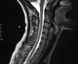
To ak stands for MRI spine and ? After the shots are received, using the special program we- view the of the switch, the radiologist makes a visual survey evaluation and forms an expert opinion. The results are carried out on photo paper, X-ray -film, on digital media, is given to the patient's hands. The pictures and the conclusion of a specialist are also brought into the history of disease.
The patient will know the results within 1-2 hours. In an individual interview, the radiologist can hold a consultation, point out the pathology found, special moments, tell you what to look for and to which specialist to seek help. After the images are examined, the radiologist interprets the result, draws up the conclusion, presents it to the treating doctor.
If the patient requires additional research, the cause is explained by the directly treating the physician.
Transcription of the MRI of the abdominal cavity
MRI of the abdominal cavity allows to assess the condition of internal organs, determine the spread and nature of the pathogen process. At the same time, it is possible to establish the diagnosis accurately and start treatment in time, preventing the development of any complications. MRI allows you to track the size of tumors in oncology. To detect timely metastases, new lesions, to make a plan for surgical intervention.
The interpretation of MRI is the most important factor on which the accuracy of the diagnosis depends. This process is quite complicated, and the as the MRI of the of the abdominal cavity is decoded, knows exactly the specialist who has deep medical knowledge of human anatomy, the characteristics of the disease. Radiologist usually has the experience of decoding on ultrasound and x-ray- devices , passes, in addition, special training courses.
If there are controversial, complex cases during decryption, several interpr of the images are used. Currently, other specialists can be remotely attracted. There is an "second opinion" service. Large medical portals on the Internet attract leading experts from all Ro ssii , as well as from abroad. The importance of such a project is obvious, especially for small towns, where the shortage of specialists is very appreciable.
Differences between MRI and CT
The stands for MRI and CT and how these survey types differ.
CT( computed tomography) - this examination is carried out using X-rays. About the difference from of conventional X-ray examination, where is two-dimensional with the image is displayed on a plate or film - n When computed tomography the image leaves volumetric. is that the device is designed so that the person is inside the device in the ring-shaped contour. Snapshots of organs are made at different angles, from different points, images are processed and a three-dimensional picture is obtained.
The principle of obtaining images on an MRI( magnetic resonance imaging) is .The difference - in the nature of the waves. The and MRI use electromagnetic waves.
The main differences between the surveys in the rays, as well as the fact that each method in its own way better reveals this or that pathology.