Computer tomography of teeth: the features of the diagnosis, the advantages of the method
Not so long ago the most effective way to get an idea of the state of bone tissue in the oral cavity area was a two-dimensional panoramic image. Currently, dentists are increasingly using computed tomography of teeth. The method allows to form voluminous images of the jaw for the expert to view the visible and hidden tissue sites from all sides.
Principle of research
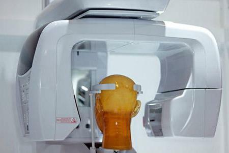 Computed tomography of teeth is based on differences in the passage of X-rays through individual tissue structures( bones, muscles).During the study, the radiation flows through the human body, after which it is captured at the output by a special detector. The result is the acquisition of a whole series of images, on the basis of which the computer 3D tomography of teeth is built.
The generated 3D model is stored in the computer memory. Later such diagnostic results can be used by other physicians in the development of therapy methods.
Diagnostic capabilities of
The use of innovative CT devices facilitates the following studies in the field of dentistry and maxillary surgery:
- Detection of defects in teeth and anomalies in the structure of bone tissue.
- Formation of ideas about the nature of focal infections.
- Collect information to prepare for surgery.
- Control over the growth of pathological tissue in the maxillofacial area.
Areas of application
 Computed tomography of teeth is indispensable primarily in surgical dentistry. The application of the research method opens the possibility for carrying out complex implantation of teeth, performing bone grafting, diagnosing tumors.
With regard to periodontology, here the computed tomography of the teeth promotes the detection of tumor-like and sclerotic defects of periodontium. Using the method, specialists also determine the level of bone resorption.
In the field of orthodontology, computed tomography of the teeth( photo tomographs are presented in this material) makes it possible to find out what condition the hard and soft tissues are in, to form an idea of the curvature of the dentition in preparation for performing the prosthesis.
Obtaining voluminous, detailed images eliminates errors in the diagnosis. The effectiveness and safety of the method makes it possible to produce therapy in a short time.
How is computerized tomography of teeth?
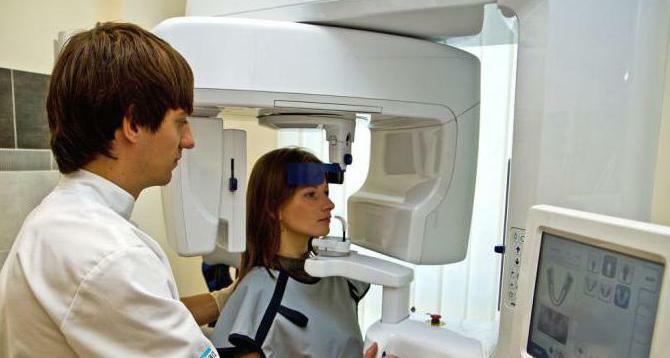
 The process of forming volumetric images with the help of modern CT apparatus is a simple, quick and painless procedure. First, the patient needs to find out where the computer tomography of the teeth is conducted in Moscow, the addresses of medical institutions that have similar equipment. With regard to the actual conduct of the study, it is performed as follows:
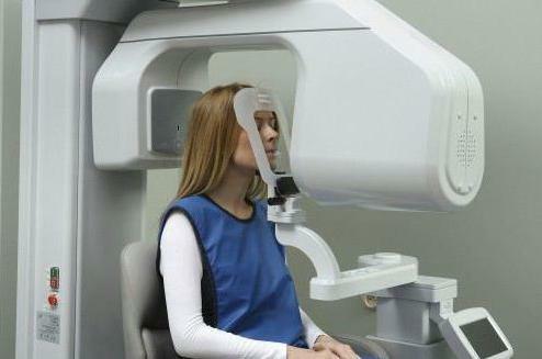
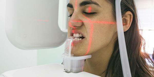
- The patient places the chin on the stand of the CT apparatus. The head is fixed in a static position. During the picture, the patient is asked to remain completely calm and do not move. Such preparation for diagnosis excludes obtaining poor-quality images.
- The specialist activates the CT apparatus. After a few moments on the monitor connected to the scanner, the first images are formed.
- When performing the diagnostics, the doctor monitors the patient's condition. The equipment is turned off as soon as the device produces such a number of images, which will suffice to create a three-dimensional model.
As a rule, computed tomography of teeth lasts no more than a minute. During this time the patient does not have time to experience discomfort. Among other things, the degree of short-term irradiation is so small that a person's health is not critically harmed.
Contraindications
 Who is not recommended for computer tomography of the teeth? Despite the general safety of the method, X-rays still affect the tissues and organs during the study. Therefore, it is necessary to take a decision on the procedure individually.
Discard study recommended:
- Pregnant women - even minimal exposure can potentially harm the unformed fetus.
- Breastfeeding mothers - X-rays have a negative effect on milk structure. In the case of a study, it is recommended that women refrain from breastfeeding for several days.
- To people who suffer from claustrophobia - to cause panic states in such patients can not only have limited space in the diagnostic room, but also rotation of the mobile part of the device, with which the volumetric images are formed.
- Patients who are difficult to maintain complete immobility for physiological reasons.
- Allergies and people with diabetes. The need to introduce a contrast medium into the body for taking pictures in this case can cause a whole lot of unpleasant consequences and harm the health.
In conclusion
 Apparently, computed tomography of teeth is an extremely effective, safe method of diagnosis, which excludes the admission of medical error. The possibility of creating a three-dimensional model of the investigated area makes it possible not only to form an idea of the state of deep bone tissue, but also to reveal various pathologies at early stages of development.