Pharynx is. .. Definition, structure and functions of pharynx, anatomical and physiological features
The pharynx is a funnel-shaped muscular canal that has a length of up to 14 cm. The anatomy of this organ allows the food lump to flow unhindered into the esophagus, and then into the stomach. In addition, thanks to anatomical and physiological characteristics, air through the pharynx from the nose to the lungs and back. That is, the digestive and respiratory system of a person crosses in the throat.
Anatomico-physiological features of
The upper part of the pharynx is attached to the base of the skull, occipital bone and temporal pyramidal bones. At the level of the 6th-7th vertebrae, the pharynx passes into the esophagus.
Inside it is a cavity( cavitas pharyngis).That is, the pharynx is the cavity.
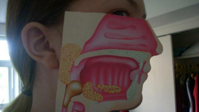
The organ is located behind the oral and nasal cavities, anterior to the occipital bone( its basilar part) and cervical upper vertebrae. In accordance with the ratio of the pharynx to other organs( that is, with the structure and functions of the pharynx), it is conditionally divided into several parts: pars laryngea, pars laryngea, pars nasalis. One of the walls( upper), which is due to the basis of the skull, is called the vault.
The nasal part of
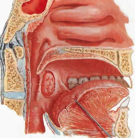
Pars nasalis in functional terms is the respiratory tract of the human pharynx. The walls of this department are immovable and therefore do not subside( the main difference from other parts of the body).
In the anterior wall of the pharynx are located the choana, and on the lateral surfaces - the pharyngeal funnel-shaped openings of the auditory tube, which is the component of the middle ear. From behind and above this hole is limited by a tube roller, which is formed by protrusion of the cartilage of the auditory tube.
The boundary between the posterior and upper walls of the pharynx is occupied by the accumulation of lymphoid tissue( on the midline) called adenoids, which in the adult are little expressed.
Between the soft sky and the opening( pharyngeal) of the tube, there is another cluster of lymphatic tissue. That is, at the entrance to the throat there is a practically dense ring of lymphatic tissue: lingual tonsil, palatine tonsils( two), pharyngeal and tubal( two) tonsils.
Oral part
Pars oralis is the middle section in the pharynx, in front communicating with the medulla of the mouth with the oral cavity, and the posterior part of it is located at the level of the third cervical vertebra. Functions of the oral part are mixed, due to the fact that the digestive and respiratory systems cross here.
Such a cross is a characteristic of the human respiratory system and was formed during the development of respiratory organs from the primary( its walls).From the rhinoceros primary bay, the oral and nasal cavities formed, the latter at the top and slightly dorsal with respect to the oral cavity. Trachea, larynx and lungs developed from the ventral( anterior) gut wall. That is why the head section of the gastrointestinal tract is located between the nasal cavity( above and dorsally) and the respiratory route( ventrally), which explains the cross of the respiratory and digestive systems in the pharynx region.
The gyrus part of
Pars laryngea is the lower part of the organ, located behind the larynx and goes from the beginning of the larynx to the beginning of the esophagus. The larynx is located on the front wall.
The structure and function of the pharynx
The basis of the pharyngeal wall is the fibrous membrane that is attached to the skull base of the skull, inside is lined with mucous membrane, and on the outside - by a muscular membrane. The latter is covered with a thin fibrous tissue that unites the pharyngeal wall with neighboring organs, and from above, it switches to m.buccinator and turns into its fascia.
The mucous membrane is covered in the pharyngeal nostril with ciliated epithelium, which corresponds to its respiratory function, and in the underlying parts it is flat multi-layered epithelium, due to which the surface becomes smooth and the food lump slips easily when swallowed. In this process, the glands and muscles of the pharynx also play a role, which are located circularly( the throats) and longitudinally( dilators).
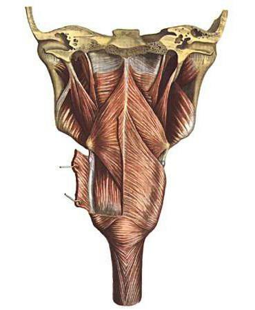
The circular layer is more developed and consists of three compressors: an upper constrictor, an average constrictor and a lower constrictor of the pharynx. Starting at different levels: from the bones of the base of the skull, the lower jaw, the root of the tongue, the cartilage of the larynx and the hyoid bone, the muscular fibers are directed back and, united, form a suture groove along the median line.
Fibers( lower) of the lower constrictor are connected with muscular fibers of the esophagus.
Muscle fibers longitudinal form the following muscles: shillopharyngeal( M. stylopharyngeus) originates from the styloid process( part of the temporal bone), runs downward and divides into two beams, enters the pharyngeal wall, and is attached to the thyroid cartilage( its upper edge);the pharyngeal muscle( M. palatopharyngeus).
Swallowing act
Due to the presence of a digestive and respiratory tract in the throat, the body is equipped with special devices that separate the respiratory tract from the digestive during swallowing. Due to the contraction of the muscles of the tongue, a lump of food is pressed against the sky by the back of the tongue and then pushed into the pharynx. At this time, the soft sky is drawn upwards( thanks to the contractions of the muscles tensor veli paratini and levator veli palatini).So the nasal( respiratory) pharynx section is completely divided with the oral department.
Together with this, the muscles that are located above the hyoid bone, pull the laryn up. The root of the tongue then descends and presses on the epiglottis, so that the latter descends, closing the passage into the larynx. After there are successive contractions of constrictors, due to which a lump of food penetrates to the esophagus. In this case, the longitudinal muscles of the pharynx work as lifters, that is, they lift the pharynx towards the movement of the food lump.
Blood supply and innervation of the pharynx
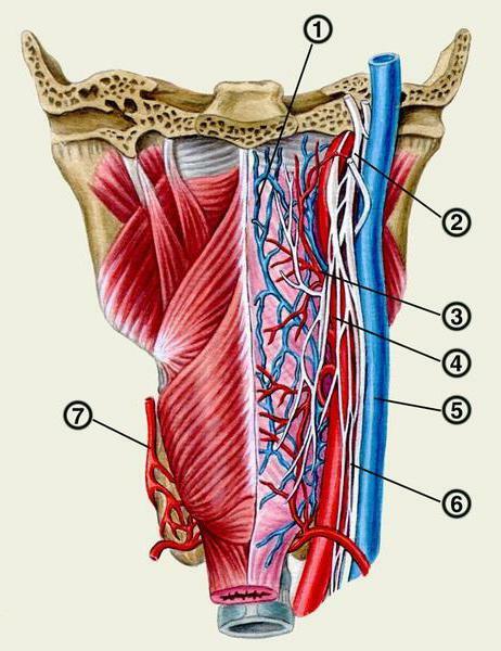
Blood supply to the pharynx mainly from the pharyngeal ascending artery( 1), the thyroid upper( 3) and branches of the facial( 2), maxillary and carotid outer arteries. Venous outflow occurs in the plexus that is located on top of the pharyngeal muscular membrane, and further along the pharyngeal veins( 4) into the jugular inner vein( 5).
Lymph flows into lymphatic cervical nodes( deep and behind-the-mouth).
The pharyngeal innervation is mediated by the pharyngeal plexus( plexus pharyngeus), which is formed by the branches of the vagus nerve( 6), the sympathetic sivola( 7), and the glossopharyngeal nerve. Sensitive innervation in this case passes through the glossopharyngeal and vagus nerves, the exception is only the shillotoch muscle, the innervation of which is carried out only by the glossopharyngeal nerve.
Dimensions
As mentioned above, the pharynx is a muscle tube. Its largest transverse dimension is at the levels of the nasal and oral cavities. The size of the pharynx( its length) averages 12-14 cm. The transverse size of the organ is 4.5 cm, that is, more than anterior-posterior size.
Diseases 
All diseases of the pharynx can be divided into several groups:
- Inflammatory acute pathology.
- Injuries and foreign bodies.
- Chronic processes.
- Disorders of the tonsils.
- of the Angina.
Inflammatory acute processes of
Among the inflammatory diseases that occur acute, we can distinguish the following:
- Pharyngitis acute - lesion of lymphoid tissue of the pharynx due to the multiplication of viruses, fungi or bacteria in it.
- Candidiasis of the pharynx - damage to the mucous organ by fungi of the genus Candida.
- Tonsillitis acute( sore throat) is the primary damage to the tonsils, which is of an infectious nature. Angina can be: catarrhal, lacunar, follicular, ulcerative-film.
- Abscess in the root of the tongue - purulent tissue damage in the area of the hyoid muscle. The cause of this pathology is infection of wounds or as a complication of inflammation of the lingual tonsil.
throat injuries Among the injuries most common are:
1. Various burns caused by electrical, radiation, thermal or chemical effects. Thermal burns develop due to getting too hot food, and chemical - when exposed to chemical agents( more often acids or alkalis).There are several degrees of tissue damage with burns:
- The first degree is characterized by erythema.
- The second degree - the formation of bubbles.
- Third degree - necrotic tissue changes.
2. Foreign bodies in the pharynx. These can be bones, pins, pieces of food and so on. The clinic for such damage depends on the depth of penetration, localization, and the size of the foreign body. There are often stitching pains, and then pain when swallowing, coughing, or a feeling of suffocation.
Chronic processes
Among the chronic lesions of the pharynx are often diagnosed:
- Pharyngitis chronic is a disease characterized by lesions of the pharyngeal mucosa of the posterior wall and lymphoid tissue as a result of acute or chronic damage to the tonsils, paranasal sinuses and so on.
- Pharyngomycosis - damage to the tissues of the pharynx caused by yeast-like fungi and developing against the background of immunodeficiencies.
- Tonsillitis chronic - autoimmune pathology of palatine tonsils. In addition, the disease is allergic-infectious and is accompanied by a persistent inflammatory process in the tissues of the palatine tonsils.