X-ray and fluorography: what's the difference, description
In this article we will consider X-rays and fluorography. What is the difference between them? At present, in our country, everyone should undergo a scheduled fluorographic examination once a year. This procedure is generally accepted and does not cause any doubt among people. However there are such situations when physicians suggest to the patient to pass a roentgen instead of a fluorography. What is more harmful - x-rays or fluorography?

Basic concepts of
Fluorography is a method of X-ray diagnostics that consists in displaying on the film the shadow of the chest organs( an outdated method) or its translation into a digital image. In turn, lung X-rays are a technique by which pathological changes are diagnosed by fixing objects onto a film. The difference between these types of X-rays is considerable. Digital fluorography is characterized by a reduced radiation effect on the patient, but its resolution is lower than the direct projection of lung radiography.
What is fluorography?
Every year, every person faces fluorography, conducted for preventive purposes. Such a procedure is carried out in medical institutions, since this is a legitimate method for screening lung pathologies. Doctors without him will not sign a commission. Fluorography has become widespread in our country due to numerous cases of tuberculosis. To prevent mass infection, the Ministry of Health came to a decision on the introduction of mandatory annual fluorography. A single dose for one study is not more than 0.015 mSv, while a preventive dose of 1 mSv is allowed. In view of this norm, it can be calculated that in order to exceed the radiation load, it is required to perform a thousand studies per year. What to choose a roentgen and a roentgenography? What is the difference between them, is of interest to many.
Types of fluorography
Currently, there are several modern versions of fluorography, which are used not only for the diagnosis of tuberculosis, but also for pneumonia.
Digital fluorography is a modern method of X-ray screening of lung diseases. This method assumes that the shadow image is photographed on a computer monitor from a special chip that is installed in the receiver. The reduced radiation load on the patient is determined by the principle of the device functioning: the ray passes through the entire area of the study in turn, after which the image is reconstructed in the software. This is what happens in the office of fluorography.
An obsolete method is traditional fluorography. With this method, the image is displayed on a small film. Thanks to this approach, cabinets were provided with high capacity, but the radiation loads were not reduced compared to pulmonary radiography.
A significant disadvantage of the digital view is considered to be the high cost of the necessary equipment, which is why not all medical facilities currently can afford such technologies. So, x-rays and fluorography - what's the difference? To understand this, you need to consider each method of diagnosis in detail.
X-ray of the lungs: what is it?
To some extent, lung X-ray is an alternative to fluorography, which has high quality, because it differs in resolution. On the pulmonary radiograph, shadows equal to two millimeters are distinguished, while in the fluorographic study the minimum size is five millimeters. Radiography is carried out with suspicion of lung diseases: pneumonia, tuberculosis, cancer and others. Children usually do not get fluorography. It is a preventive method.
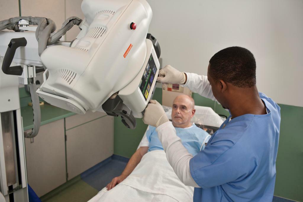
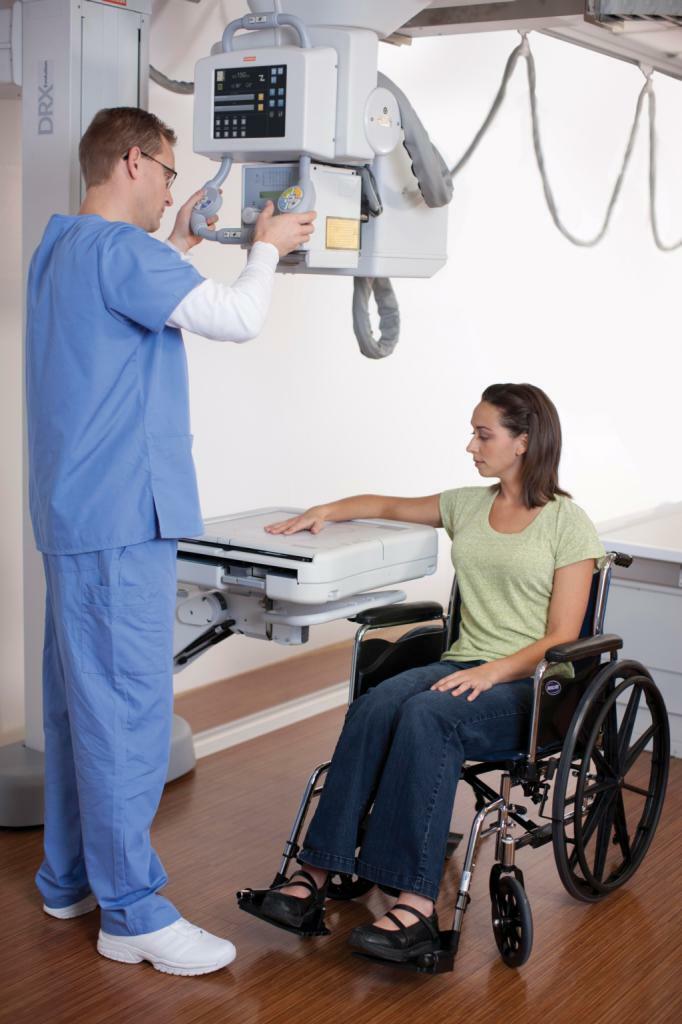
X-ray images are obtained by exposing certain areas of the film as X-rays pass through the body. How do X-rays do? About this below.
Is there any danger?
During the study, a high, but non-long-term radiation load on a person is formed. The danger of it is that there may be mutations at the cellular level. That is why, before sending a patient to a radiograph, the attending physician should compare the degree of risk from x-ray exposure with the practical value of the results obtained during the examination. The procedure is assigned when this value is low. X-ray diagnostics is based on the principle: the benefit must exceed the harm.
This must be remembered when a tooth X-ray is administered during pregnancy. Do it only in the most extreme cases.
Radiographic safety of WGC
It should be said that the magnitude of the radiation load on a patient with lung X-ray in domestic medical institutions is higher than that of developed countries. This is because old equipment is used. For example, in Europe with X-ray study, the average dose of one patient per year is not more than 0.6 mSv. In our country, it is twice as high - about 1.5 mSv. For greater safety, it is recommended to do diagnostic work on an X-ray machine in modern institutions. Of course, if acute pneumonia is diagnosed, the doctor is limited in time and will not allow the patient to choose a clinic for examination.
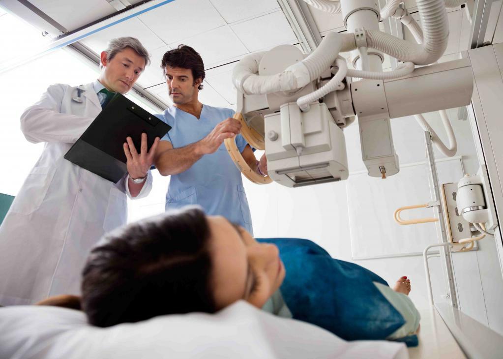
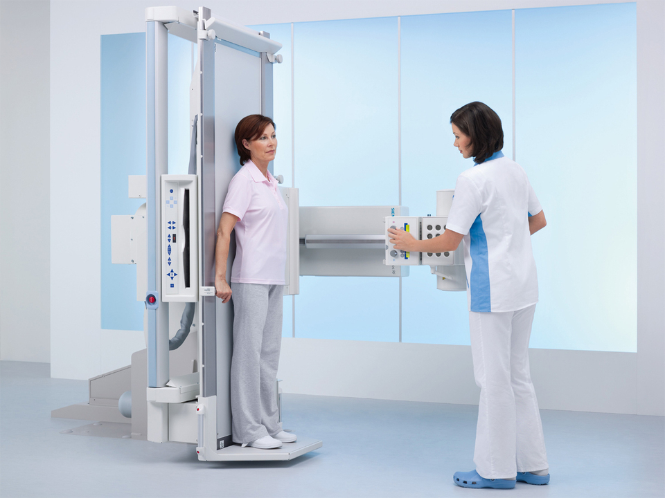
In this case, pathology poses a threat to life, so that what is available will be used for analysis. In this situation, a snapshot of the lungs will be performed, not just in a direct projection, but also in the side, and perhaps also in the sighting. This is required in order to determine the size, as well as the prevalence of the focus of pathology in the lung tissue. There are such important contraindications to fluorography and X-rays, as planning a child and pregnancy. When is a medical X-ray necessary? Indications for the radiography of WGCs, that is, chest organs, are the suspicions available to the doctor of the pathology of the lungs( cancer, tuberculosis, pneumonia).Special training is not required. There is only one condition - to remove foreign objects and to expose the thorax. Shooting can also be carried out in underwear if there are no metal objects and synthetic fibers that can be reflected on the roentgenogram. The transparency of the upper parts of the pulmonary fields in women can decrease if they are closed during the procedure by the hair. This feature is taken into account by the radiologist during the analysis of the image.
Types of
There are the following types of pulmonary radiography:
- sighting;
- review.
During the targeted examination, the focus on a particular pathological tissue site occurs. Aiming images on the X-ray machine should be done under control, however, there is an increase in radiation exposure to the patient. With the survey methodology, it is necessary to take pictures in two projections: the lateral and the direct. The main cause of errors that may appear in the picture is in the dynamic blurriness, that is, fuzzy contours of the formations caused by pulsation of large vessels or breathing. It can be eliminated when the exposure time is 0.02 to 0.03 seconds.
That's why experts recommend taking light shots at exposures from 0.1 to 0.15 seconds. Of course, in this case, you need powerful equipment. To prevent projective distortion, the distance between the focus and the object should be from one and half to two meters. What is better - to visit the office of fluorography or X-ray?
Fluorography or X-ray: what is better with pneumonia?
Often, patients are interested in whether it is possible to refuse a lung X-ray or from fluorography? By law, a person has this right, but at the same time he is responsible for his own health. If the refusal is written, then the medical commission can be passed, however the phthisiatrician can not sign it, since he has every right. If a specialist has suspicions of pneumonia or active tuberculosis, as well as confirmation of these pathologies with other clinical and instrumental methods( increase in white blood cells, sputum analysis), the doctor can, by law, refer the patient to compulsory treatment.

Danger of tuberculosis
Tuberculosis in an open form is dangerous for surrounding people, and therefore it should be treated in TB hospitals. A threat to life is also pneumonia, clearly manifested in the pulmonary radiograph. There are no other reliable methods for its detection. Fluorography is not carried out for children, it costs x-rays.
The presence of processes of inflammation in the lung tissue and the appointment of antibiotics can be based on indirect signs, but with a full-fledged radiographic analysis it is possible to monitor the extent, size of the foci, the severity and course of the pathology process. The doctor can combine several antibacterial agents and change the treatment plan during an exacerbation. If you require a fluorographic coupon at a dentist, ophthalmologist or other specialists, the actions of medical workers are illegal, because internal orders are not able to cancel the constitutional action. You just need to write a refusal in your outpatient card or a medical history about the failure of such an investigation. When deciding what is best done - chest X-ray and fluorography, it is necessary to evaluate the specificity of both methods and their preventive use in setting the diagnosis.
The expediency of conducting a lung X-ray or fluorography is actively discussed by researchers, scientists and the media. Each person may have their own opinion, but the best way to choose the method of X-ray examination is to rely on the opinion of the doctor, since it is necessary to take into account the relationship between the practical benefit and the harm produced by ionizing radiation.
Negative effects of
Fluorography and radiography adversely affect the human body. The degree of X-ray dose control is 1.5 mSv per gram. With film fluorography, this indicator varies from 0.5 to 0.8 mSv, for the digital one - 0.04.For the examination of organs located in the chest, the level of EED is required. When performing an examination by means of an X-ray machine, the image appears on a special film. During fluorography, the preliminary image is displayed on the monitor, after which it is photographed. Thanks to this technique, pathology can be diagnosed. The rays pass through the body through the X-ray, reflecting on the film.
Another technique is characterized by additional transformation of rays into pronounced light. After that, the image in a reduced form is focused on the film. Based on its results, an additional examination is carried out. That is why x-rays or fluorography are assigned individually in each case. Radiography of OGC is used for screening of lungs and tuberculosis. For this purpose, stationary and mobile equipment are used. It is better not to prescribe the X-ray of a tooth during pregnancy.
In medicine, now the digital technique replaces the film technique, because it greatly facilitates the work with the image. On the screen of the monitor, a snapshot is displayed, printed out and then transmitted over the network, and then downloaded to the database. Such a survey is characterized by a reduced radiation load and a low expenditure on materials.

Now we know that it shows X-rays, and that fluorography.
Main findings of
We examined various methods of radiographic research. During the radiography on a special film, an image is displayed, and with fluorography it is reflected on the screen, and from there it is photographed on a digital or ordinary camera. With fluorography, the radiation load is higher in comparison with radiography. Most often, fluorography is used to diagnose diseases, and x-rays are used to refine or monitor the pathology in the dynamics. The first method has a lower cost.
We examined X-rays and fluorography. What is the difference between them, now readers know.