Auditory ossicles: general structure
The human ear is a unique organ functioning on a pair basis, which is located in the very depth of the temporal bone. The anatomy of its structure allows to catch mechanical fluctuations of air, and also to carry out their transfer on internal environments, then to transform a sound and transfer it to the brain centers.
According to the anatomical structure, human ears can be conditionally divided into three parts, namely the outer, middle and inner. 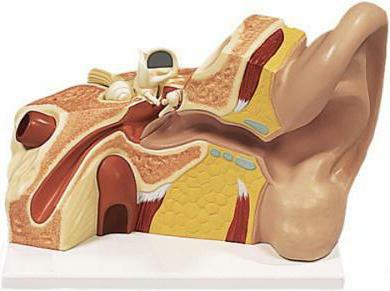
Middle ear elements
Studying the structure of the middle part of the ear, you can see that it is divided into several parts: the drum cavity, the ear tube and the auditory ossicles. The latter include an anvil, a hammer and a stirrup.
Hammer of the middle ear
This part of the auditory ossicles includes such elements as the neck and handle. The head of the malleus is connected by means of a hammer joint with the structure of the anvil body. And the handle of this malleus is connected with the tympanic membrane by fusion with it. To the neck of the malleus is attached a special muscle, which stretches the eardrum. 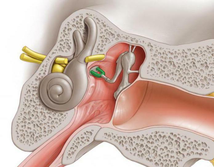
Anvil
This ear element has at its disposal a length of six to seven millimeters, which consists of a special body and two legs with short and long dimensions. The one that is short, has a lenticular process that fuses with the anvil stapler joint and with the head of the stirrup itself.
What else does the auditory ossicle of the middle ear include?
Stirrup
The stirrup has a head, as well as front and rear legs with a part of the base. To its rear leg is attached a stirrup muscle. The base of the stirrup itself is built into the oval window of the vestibule of the labyrinth. A ring binder in the form of a membrane that is located between the staple support base and the edge of the oval window contributes to the mobility of this auditory element, which is provided by the action of air waves directly on the tympanic membrane. 
Anatomical description of the muscles attached to the bones
Two transverse striated muscles are attached to the auditory bones, which perform certain functions for the transmission of sound vibrations.
One of them pulls the eardrum and originates from the walls of the muscular and tubal channels related to the temporal bone, and then it attaches to the neck of the malleus itself. The function of this tissue is to pull the hammer handle inside. The tension is toward the drum cavity. At the same time, the tympanic membrane exerts pressure and is therefore taut and concave into the region of the middle ear.
Another muscle of the stapes originates in the thickness of the pyramidal ridge of the mastoid wall of the tympanic region and is attached to the stalk of the stapes located posteriorly. Its function is to cut and remove from the hole the base of the stirrup itself. During a powerful oscillation of the auditory ossicles, along with the previous muscle, auditory ossicles are retained, which significantly reduces their displacement.
Auditory ossicles, which are interconnected by joints, and, in addition, muscles related to the middle ear, completely regulate the movement of air currents at different levels of intensity. 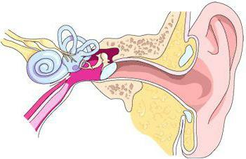
Drum cavity of the middle ear
In addition to ossicles, a certain cavity is also included in the structure of the middle ear, which is usually called a drum. The cavity is located in the temporal part of the bone, and its volume is one cubic centimeter. In this area just located auditory ossicles with a tympanic membrane nearby.
Above the cavity is located the mastoid process, which consists of cells carrying air flows. In it, there is a certain cave, that is, a cell through which the movement of air molecules takes place. In the anatomy of the human ear, this region performs the role of the most characteristic reference point in the implementation of any surgical interventions. How are connected ossicles, are interested in many.
Auditory tube in the anatomy of the structure of the human's middle ear
This area is a formation that can reach a length of three and a half centimeters, and the diameter of its lumen can be up to two millimeters. Its upper origin is located in the tympanic region, and the lower pharyngeal opening opens in the nasopharynx approximately at the level of the hard palate. 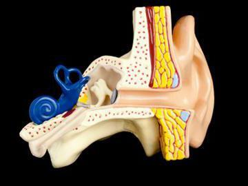
The auditory tube consists of two sections, which are separated by the narrowest place in its area, the so-called isthmus. From the drum region goes the bone part, which extends below the isthmus, it is commonly called membranous-cartilaginous.
The walls of the tube located in the cartilaginous region are usually closed in a calm state, but they can open slightly when chewing, and this can also occur during swallowing or yawning. The increase in the lumen of the tube occurs through two muscles, which are connected with the palatal curtain. The ear shell is covered with epithelium and has a mucous surface, and its cilia move to the pharyngeal mouth, which makes it possible to perform the drainage function of the tube.
Other facts about the auditory ossicle in the ear and the structure of the middle ear
The middle ear is directly connected to the nasopharynx by means of the Eustachian tube, whose immediate function is to adjust the pressure coming from the air. A sharp bookmark of the human ears can signal a rapid decline, or an increase in the pressure of the environment.
Long and prolonged tenderness in the temples, most likely, indicates that the ears are currently trying to actively fight the infection and protect the brain from all sorts of violations of its performance.
Internal auditory ossicle 
A number of fascinating facts of pressure can also include reflex yawning, which indicates that in the human environment, its sharp changes occurred, and therefore a response was induced in the form of yawning. It should also be known that the human's middle ear has a mucous membrane in its structure.
Do not forget that unexpected, even, like sharp sounds can provoke muscle contraction on a reflex basis and damage both the structure and functioning of the hearing. The functions of the auditory ossicles are unique.
All listed elements of the anatomical structure carry such a functional ability of the auditory ossicles as the transmission of perceived noise, as well as its transfer from the outer ear region to the inner one. Any violation and failure of the functioning of at least one of the structures can lead to the destruction of the work of the hearing organs completely.
Inflammation of the middle ear
The middle ear is a small cavity between the inner and outer ear. In the middle ear, the transformation of air vibrations into the oscillation of the fluid is provided, which is recorded by the auditory receptors in the inner ear. This happens with the help of special bones( malleus, anvil, stapes) due to sound vibration from the tympanic membrane to the auditory receptors. To equalize the pressure between the cavity and the environment, the middle ear is reported by the Eustachian tube with the nose. Infectious agent penetrates into this anatomical structure and provokes inflammation - otitis media.